Sassoon Road


Sassoon Road
Hinjawadi
Wanworie
Sassoon Road
Sassoon Road
Hinjawadi
Wanworie
Sassoon Road
Sassoon Road

The Department of Nuclear Medicine and Molecular Imaging, established in 1996 at Ruby Hall Clinic, was the first hospital-owned Nuclear Medicine Department in Maharashtra, located outside of Mumbai, marking a significant milestone. The Department offers a comprehensive suite of advanced diagnostic and therapeutic services designed to detect, monitor and treat a wide variety of conditions at the molecular level.
From renogram studies and bone scans to highly specialised PET imaging and targeted radionuclide therapies, the department is equipped with state-of-the-art technology, including its own in-house GE MINItrace Cyclotron, ensuring timely access to essential radiopharmaceuticals.
The department excels in both functional diagnosis and targeted treatments, offering world-class care for patients with cancer, joint diseases, thyroid conditions and more.
Our team consists of the best nuclear medicine physicians and nuclear medicine specialists, who focus on specialised nuclear medicine.
1. Renal Scan (DTPA / MAG3 Renal Scintigraphy)
A renal scan is a nuclear medicine test that assesses how well your kidneys are working. It uses a small amount of radioactive material—commonly Tc-99m DTPA or MAG3 – which is injected into the bloodstream and filtered by the kidneys. A special camera (gamma camera) tracks how the kidneys take up, process, and excrete the tracer.
This scan provides valuable information about:
– Kidney function and drainage
– Presence of any blockages or obstructions
– Comparison of function between the two kidneys
Common indications:
– Evaluating kidney function before surgery
– Suspected urinary tract obstruction
– Monitoring renal transplant function
Preparation:
– The patient may be asked to drink water before the test.
– A mild diuretic (Lasix) may be given during the scan to assess urinary drainage.
2. Radio Ablations
What is it?
This therapy uses radioactive substances to ablate (destroy) specific tissues, most commonly in thyroid or liver cancers.
Functioning:
– The radiopharmaceutical travels through the bloodstream and binds to the target tissue.
– Once bound, it emits radiation that gradually destroys the abnormal cells.
Used for:
– Residual thyroid tissue post-surgery
– Small metastatic lesions (bone/liver)
– Liver tumours (in cases where surgery isn’t possible)
Procedure and preparation are similar to high-dose I-131 therapy, depending on the radiopharmaceutical used.
3. PRRT – Peptide Receptor Radionuclide Therapy
What is PRRT?
Who may benefit from PRRT?
Why would my doctor prescribe PRRT?
How is PRRT administered?
When is PRRT administered?
There are three main PRRT types offered:
i) DOTA-PRRT (177Lu-DOTATATE)
Used for: Neuroendocrine tumours (NETs) that express somatostatin receptors
Mechanism: A somatostatin analogue labelled with Lutetium-177 targets the receptors on tumour cells and emits radiation to kill them from inside.
Procedure:
– IV infusion in cycles (4–6 weeks apart)
– Typically requires 1–2 days of hospital admission per cycle
Preparation:
– 68Ga-DOTATATE scan to confirm receptor expression
– Blood tests, kidney function, hydration before therapy
ii) PSMA-PRRT (177Lu-PSMA Therapy)
What is PRRT (Peptide Receptor Radionuclide Therapy) for Prostate Cancer?
PSMA PRRT (Peptide Receptor Radionuclide Therapy) is a targeted nuclear medicine treatment for advanced prostate cancer. It uses a small protein (peptide) that binds to the PSMA receptor—found in high levels on prostate cancer cells—linked to a radioactive substance, Lutetium-177. Once injected, the peptide targets cancer cells and delivers beta radiation directly to them, minimising damage to healthy tissue. It is especially used when the cancer has spread to bones or lymph nodes.
How is it Done?
– IV injection of Lutetium-177 attached to the PSMA-targeting peptide
– Radiation kills cancer cells by binding to PSMA receptors
– Has shown promising outcomes in advanced, metastatic prostate cancer
Used for:
Advanced prostate cancer with PSMA-positive tumours
Mechanism:
A radiolabelled molecule targets the Prostate-Specific Membrane Antigen, emitting beta radiation directly to cancer cells.
Benefits:
– Treats metastases in bones, lymph nodes, and soft tissues
– Often used when other therapies are no longer effective
Procedure:
– IV Infusion in cycles (4-6 weeks apart)
– Typically requires 1 day stay in Hospital
– 68Ga-PSMA PET required for eligibility
PREPARATION BEFORE THERAPY
Steps to Prepare for Treatment:
– Medical Review: Ensure the patient is a suitable candidate based on medical
history.
– Imaging: A ⁶⁸Ga-PSMA PET/CT scan helps assess disease spread and plan treatment.
– Blood Tests: Kidney function and overall health must be evaluated beforehand.
– Hydration: Good hydration is important to protect kidneys during treatment.
– Scheduling: Book appointments 10–12 days in advance to arrange the ¹⁷⁷Lu-PSMA dose.
– Emotional Support: Address patient and family concerns, and offer support resources.
– Follow-up: Monitor closely post-treatment to assess response and manage side effects.
About the Therapy
– Cycles: Given in 4 cycles, spaced 4–5 weeks apart, with 1–2 days hospital stay per cycle
– Mechanism: A radiolabelled molecule targets PSMA receptors on prostate cancer cells, delivering radiation directly to them
– Effectiveness: Can shrink tumours, relieve symptoms, and extend survival in advanced cases
– Monitoring: ⁶⁸Ga-PSMA PET/CT used to track response – Side Effects: Typically mild—may include fatigue, nausea, or reduced blood counts
– Tolerance: Most patients handle treatment well with minimal complications
iii) FAPI-PRRT (Emerging Therapy)
Used for:
Solid tumours where traditional PRRT is not effective, such as breast, lung, ovarian, pancreatic and gastric cancers.
How it works:
Targets fibroblast activation protein found in tumour-supporting stromal cells. Delivers radiation not only to the cancer but to its microenvironment.
Note:
Still under clinical trials in many parts of the world but shows promising results, especially when FDG or PSMA is not suitable.
4. TARE (Transarterial Radioembolization)
What is TARE?
A targeted therapy for liver tumours, both primary (like HCC) and secondary (metastatic).
How it works:
Tiny radioactive beads (Yttrium-90) are injected into the hepatic artery. These beads lodge in the tumour’s blood vessels and emit radiation directly into the tumour, sparing healthy liver tissue.
Used for:
– Hepatocellular carcinoma (HCC)
– Liver metastases from colorectal, breast and neuroendocrine tumours
Procedure:
– Performed by an interventional radiologist
– Outpatient or short stay
– Requires angiography and mapping before treatment
Preparation:
– Liver function tests
– Planning angiogram
– Imaging with 99mTc-MAA to ensure safe distribution
5. Radiation Synovectomy
What is it?
A non-surgical therapy for chronic joint inflammation (like in rheumatoid arthritis or haemophilia-related arthritis).
How it works:
A small dose of radioactive material is injected directly into the joint space. The radiation reduces inflammation and thickened joint lining (synovium).
Used for:
– Persistent arthritis in knees, ankles, elbows
– Haemophilic arthropathy
– Inflammatory arthritis unresponsive to other treatments
Procedure:
– Outpatient injection into the joint
– Often followed by joint immobilisation for 24–48 hours
Preparation:
– Blood tests to rule out infection
– Avoid NSAIDs before the procedure
– No special fasting needed
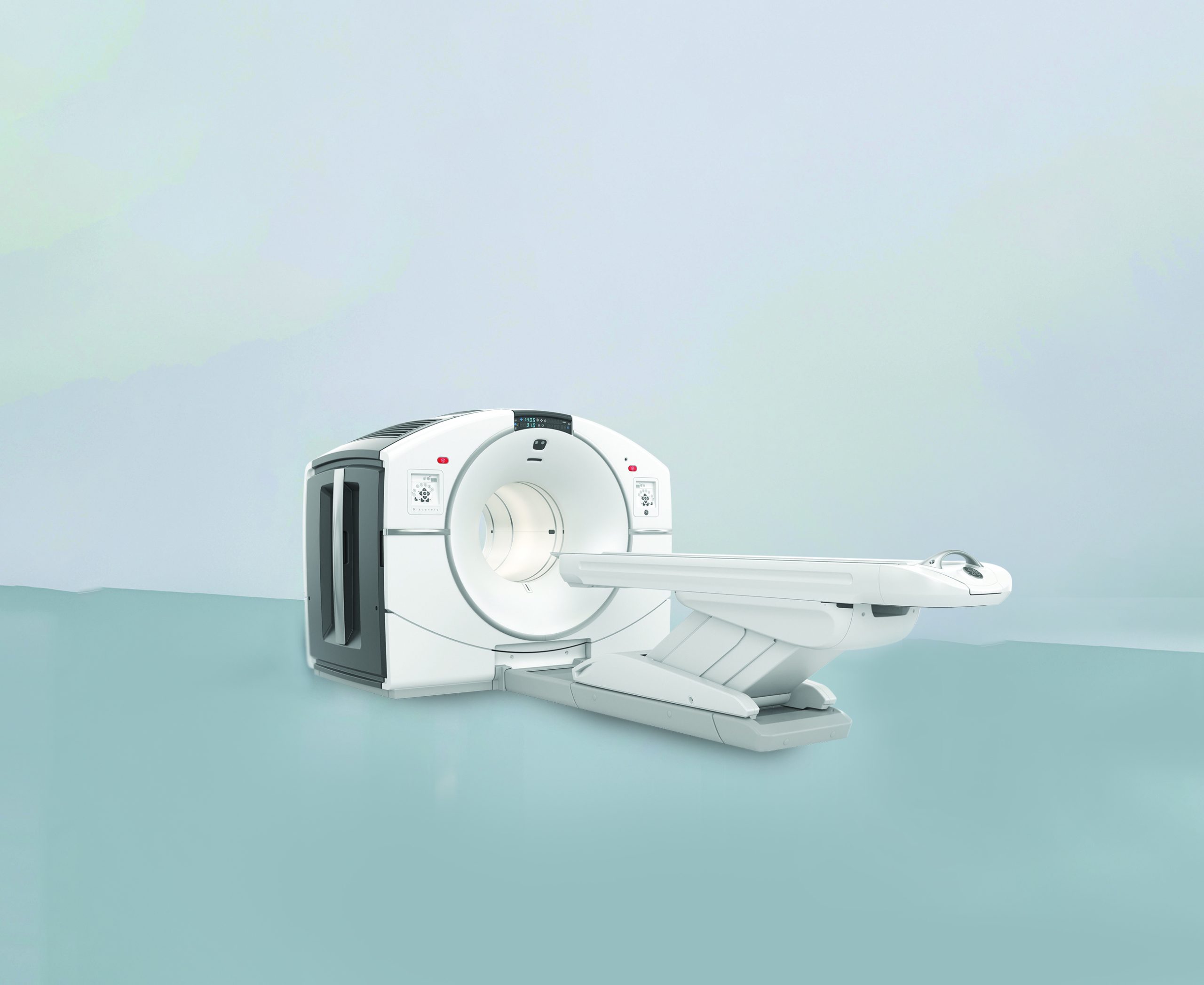
Molecular Imaging is an advanced form of nuclear medicine that provides highly detailed, functional images of what’s happening inside the body at the molecular and cellular level. PET/CT scans are commonly used in this category, combining functional imaging (PET) with anatomical detail (CT).
PET Scans (Positron Emission Tomography)
PET scans use a radioactive tracer (commonly FDG) to show metabolic activity inside the body. Cancer cells tend to be more metabolically active than normal cells, which means they absorb more tracer and appear brighter on the scan.
What it shows:
– Early detection of cancers
– Staging of cancers (how far they’ve spread)
– Treatment response evaluation
– Recurrence detection
Procedure:
– The tracer is injected intravenously.
– After a short waiting period (usually 45–60 minutes), the scan is performed.
– Patients lie still while the PET/CT camera captures images for 10-15 minutes only
Preparation:
– Fast for 4–6 hours before the scan.
– Blood sugar should be under control.
– Avoid vigorous exercise 24 hours before.
1. FDG Scans (Fluorodeoxyglucose PET Scan)
FDG is a radioactive glucose molecule used in most PET scans. It highlights areas of increased sugar metabolism, which often correlates with cancer activity.
Applications:
– Wide range of cancers: lung, breast, colon, lymphoma, etc.
– Inflammation and infection imaging
– Brain and cardiac evaluations
Patient instructions:
– Same as standard PET scan (fasting, control blood sugar)
– Diabetic patients require special scheduling
2. DOTA Scans (68Ga-DOTATATE PET Scan)
DOTA scans use Gallium-68 DOTATATE, which binds to somatostatin receptors found in neuroendocrine tumours (NETs). This scan is highly sensitive in detecting NETs throughout the body.
What it helps with:
– Diagnosis and staging of neuroendocrine tumours
– Assessing eligibility for DOTA PRRT therapy
– Monitoring therapy response
Procedure:
– IV injection of 68Ga-DOTATATE
– Scan performed after 45–60 minutes
– Entire process takes 1.5–2 hours
Preparation:
– No major dietary restrictions
– Routine medications can usually be continued
3. PSMA Scans (⁶⁸Ga-PSMA PET Scan)
PSMA PET/CT is a highly accurate molecular imaging technique for detecting prostate cancer, especially when conventional methods (CT, MRI, bone scan, or even FDG PET/CT) fall short. Unlike other cancers, prostate cancer cells use very little glucose, making standard PET scans less effective. PSMA PET/CT uses a molecule that targets the Prostate-Specific Membrane Antigen (PSMA) on prostate cancer cells, combined with Gallium-68 for precise imaging.
Why Choose ⁶⁸Ga-PSMA?
– Tumour-specific: Directly detects prostate cancer cells via PSMA, not just indirect signs like bone changes or lymph node enlargement.
– High sensitivity: Detects small regional and distant metastases with excellent tumour-to-background contrast.
– Accurate recurrence localisation: Useful even at low PSA levels post-treatment.
– Anatomical correlation: Integrated CT scan enhances diagnostic accuracy.
Uses:
– Detection of prostate cancer recurrence even at low PSA levels
– Staging of newly diagnosed prostate cancer
– Treatment planning for PRRT
– Lutetium-177-labeled anti-prostate-specific membrane antigen monoclonal antibody for metastatic, castration resistant prostate cancer, is also available for suitable patients.
Preparation:
– Mild hydration
– Minimal fasting (2–4 hours before scan)
– No special medication restrictions unless advised
4. FAPI Scans (Fibroblast Activation Protein Inhibitor PET Scan)
FAPI PET is a newer imaging technique that targets fibroblast activation protein, often present in the tumour microenvironment. This scan can detect tumours that do not show up well on FDG PET, including some breast, ovarian, lung & oesophageal cancers.
Advantages:
– Better imaging for cancers with low metabolic activity
– Less background noise than FDG
– Can be used when FDG PET is inconclusive
Procedure:
– Similar to FDG or PSMA PET scan in timing and comfort
– Still under study in many centres but rapidly gaining ground
Preparation:
– Minimal preparation; fasting may be required based on protocol
5. Exendin Scan (⁶⁸Ga-Exendin PET Scan)
Exendin scans use Gallium-68 labelled Exendin, which targets GLP-1 receptors predominantly found in insulinomas. This highly specialised scan is particularly useful for detecting small or elusive insulin-secreting tumours.
What it helps with:
– Localisation of insulinomas (especially small or occult lesions)
– Differentiating insulinoma from other pancreatic lesions
– Surgical planning and preoperative evaluation
Procedure:
– IV injection of ⁶⁸Ga-Exendin
– Scan performed after 60–90 minutes
– Entire process takes approximately 2–2.5 hours
Preparation:
– Fasting for at least 6 hours prior to scan
– Blood sugar monitoring before and during the procedure
– Routine medications may need adjustment (especially anti-diabetics; consult
physician)

Nuclear medicine therapies use targeted radioactive substances to treat conditions, primarily cancers, by delivering radiation directly to tumour cells while sparing most healthy tissue.
High Dose Therapy Facility
These therapies may be used alongside surgery or chemotherapy. A radioactive drug (given orally or via IV) travels through the body, attaches to cancer cells, and destroys them with radiation. Unbound drug is excreted, mainly through urine.
What is Theranostics?
A fast-growing field in nuclear medicine, Theranostics uses specific radioisotopes to both detect and treat tumour cells with minimal harm to normal tissue.
Available Theranostic Treatments:
– Thyroid cancer: Treated with oral radioactive iodine post-surgery or in metastatic cases
– Neuroendocrine, prostate, and medullary cancers: Treated with IV radioisotopes
– Liver cancers and metastases: Treated with targeted radioisotope injections
– Bone metastases (pain relief): IV therapy for prostate cancer patients
– Joint conditions: Intra-articular therapy for synovitis and haemophilia
– TARE (90Yttrium): For liver tumours—our centre pioneered this intra-hepatic embolisation
What to Expect:
– Fasting from midnight before treatment
– Temporary pause of thyroid medications (if applicable)
– Post-treatment isolation in a specialised, air-conditioned room equipped with monitoring, oxygen, and ventilation, located near the ICU for added safety
1. High Dose Therapies (Radioiodine Therapy – I-131)
What is it?
High-dose radioiodine therapy involves using Iodine-131, a radioactive form of iodine, to treat thyroid-related conditions.
How it works:
I-131 is taken up naturally by thyroid tissue. When administered in high doses, it emits beta radiation that destroys thyroid cells, including cancerous or overactive cells.
Used for:
– Thyroid cancer (after surgery to remove remaining tissue or cancer spread)
– Hyperthyroidism (overactive thyroid due to Graves’ disease or toxic nodules)
Procedure:
– I-131 is given orally in capsule or liquid form.
– The patient may need to stay isolated for 1–3 days, depending on the dose.
Preparation:
– Stop certain thyroid medications beforehand.
– Follow a low-iodine diet for 1–2 weeks prior.
– No pregnancy or breastfeeding during or shortly after therapy.
2. Radio Ablations
What is it?
This therapy uses radioactive substances to ablate (destroy) specific tissues, most commonly in thyroid or liver cancers.
Functioning:
– The radiopharmaceutical travels through the bloodstream and binds to the target tissue.
– Once bound, it emits radiation that gradually destroys the abnormal cells.
Used for:
– Residual thyroid tissue post-surgery
– Small metastatic lesions (bone/liver)
– Liver tumours (in cases where surgery isn’t possible)
Procedure and preparation are similar to high-dose I-131 therapy, depending on the radiopharmaceutical used.
3. PRRT – Peptide Receptor Radionuclide Therapy
What is PRRT?
Who may benefit from PRRT?
Why would my doctor prescribe PRRT?
How is PRRT administered?
When is PRRT administered?
There are three main PRRT types offered:
i) DOTA-PRRT (177Lu-DOTATATE)
Used for: Neuroendocrine tumours (NETs) that express somatostatin receptors
Mechanism: A somatostatin analogue labelled with Lutetium-177 targets the receptors on tumour cells and emits radiation to kill them from inside.
Procedure:
– IV infusion in cycles (4–6 weeks apart)
– Typically requires 1–2 days of hospital admission per cycle
Preparation:
– 68Ga-DOTATATE scan to confirm receptor expression
– Blood tests, kidney function, hydration before therapy
ii) PSMA-PRRT (177Lu-PSMA Therapy)
What is PRRT (Peptide Receptor Radionuclide Therapy) for Prostate Cancer?
PSMA PRRT (Peptide Receptor Radionuclide Therapy) is a targeted nuclear medicine treatment for advanced prostate cancer. It uses a small protein (peptide) that binds to the PSMA receptor—found in high levels on prostate cancer cells—linked to a radioactive substance, Lutetium-177. Once injected, the peptide targets cancer cells and delivers beta radiation directly to them, minimising damage to healthy tissue. It is especially used when the cancer has spread to bones or lymph nodes.
How is it Done?
– IV injection of Lutetium-177 attached to the PSMA-targeting peptide
– Radiation kills cancer cells by binding to PSMA receptors
– Has shown promising outcomes in advanced, metastatic prostate cancer
Used for:
Advanced prostate cancer with PSMA-positive tumours
Mechanism:
A radiolabelled molecule targets the Prostate-Specific Membrane Antigen, emitting beta radiation directly to cancer cells.
Benefits:
– Treats metastases in bones, lymph nodes, and soft tissues
– Often used when other therapies are no longer effective
Procedure:
– IV Infusion in cycles (4-6 weeks apart)
– Typically requires 1 day stay in Hospital
– 68Ga-PSMA PET required for eligibility
PREPARATION BEFORE THERAPY
Steps to Prepare for Treatment:
– Medical Review: Ensure the patient is a suitable candidate based on medical
history.
– Imaging: A ⁶⁸Ga-PSMA PET/CT scan helps assess disease spread and plan treatment.
– Blood Tests: Kidney function and overall health must be evaluated beforehand.
– Hydration: Good hydration is important to protect kidneys during treatment.
– Scheduling: Book appointments 10–12 days in advance to arrange the ¹⁷⁷Lu-PSMA dose.
– Emotional Support: Address patient and family concerns, and offer support resources.
– Follow-up: Monitor closely post-treatment to assess response and manage side effects.
About the Therapy
– Cycles: Given in 4 cycles, spaced 4–5 weeks apart, with 1–2 days hospital stay per cycle
– Mechanism: A radiolabelled molecule targets PSMA receptors on prostate cancer cells, delivering radiation directly to them
– Effectiveness: Can shrink tumours, relieve symptoms, and extend survival in advanced cases
– Monitoring: ⁶⁸Ga-PSMA PET/CT used to track response – Side Effects: Typically mild—may include fatigue, nausea, or reduced blood counts
– Tolerance: Most patients handle treatment well with minimal complications
iii) FAPI-PRRT (Emerging Therapy)
Used for:
Solid tumours where traditional PRRT is not effective, such as breast, lung, ovarian, pancreatic and gastric cancers.
How it works:
Targets fibroblast activation protein found in tumour-supporting stromal cells. Delivers radiation not only to the cancer but to its microenvironment.
Note:
Still under clinical trials in many parts of the world but shows promising results, especially when FDG or PSMA is not suitable.
4. TARE (Transarterial Radioembolization)
What is TARE?
A targeted therapy for liver tumours, both primary (like HCC) and secondary (metastatic).
How it works:
Tiny radioactive beads (Yttrium-90) are injected into the hepatic artery. These beads lodge in the tumour’s blood vessels and emit radiation directly into the tumour, sparing healthy liver tissue.
Used for:
– Hepatocellular carcinoma (HCC)
– Liver metastases from colorectal, breast and neuroendocrine tumours
Procedure:
– Performed by an interventional radiologist
– Outpatient or short stay
– Requires angiography and mapping before treatment
Preparation:
– Liver function tests
– Planning angiogram
– Imaging with 99mTc-MAA to ensure safe distribution
5. Radiation Synovectomy
What is it?
A non-surgical therapy for chronic joint inflammation (like in rheumatoid arthritis or haemophilia-related arthritis).
How it works:
A small dose of radioactive material is injected directly into the joint space. The radiation reduces inflammation and thickened joint lining (synovium).
Used for:
– Persistent arthritis in knees, ankles, elbows
– Haemophilic arthropathy
– Inflammatory arthritis unresponsive to other treatments
Procedure:
– Outpatient injection into the joint
– Often followed by joint immobilisation for 24–48 hours
Preparation:
– Blood tests to rule out infection
– Avoid NSAIDs before the procedure
– No special fasting needed
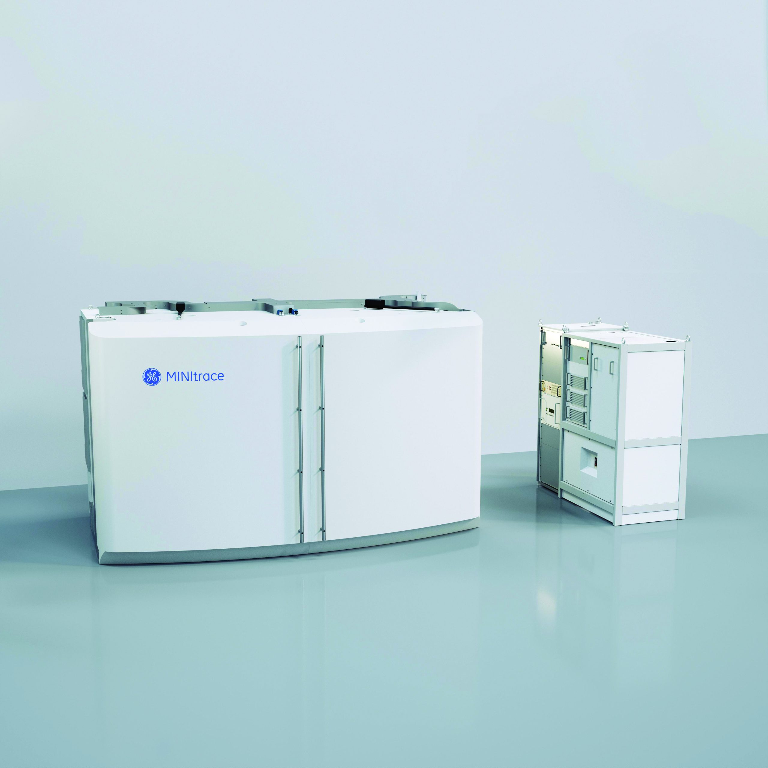
What is a Cyclotron?
A cyclotron is a particle accelerator used in nuclear medicine to produce radioisotopes for PET scans and targeted therapies. The GE MINItrace Cyclotron, Pune’s first, enables on-site production of high- quality radiotracers, improving diagnostic accuracy and treatment speed.
Key Benefits:
• On-site radiotracer production for quicker, more efficient PET/CT Scans
• Enhanced imaging precision for accurate diagnosis
• Faster turnaround, reducing waiting times and improving patient experience
• Supports theranostics for targeted treatment approaches
• Broader diagnostic applications for comprehensive disease management
How it Works:
– Accelerates protons to collide with target material
– Produces radioactive isotopes
– Isotopes are synthesised into radiopharmaceuticals for scans or treatment
What Does It Produce?
The GE MINItrace Cyclotron can produce a wide variety of PET radioisotopes, including:
– 18F-FDG (Fluorodeoxyglucose) – for detecting metabolic activity (most common PET tracer)
– 68Ga (Gallium) – used in PSMA and DOTA scans
– 13N – for specialised imaging (neuro, cardiac, oncology)
– FAPI tracers – under research and specialised production
Why it Matters?
– Enables real-time availability of short-lived isotopes
– Ensures better image resolution and faster scans
– Prepares custom doses for therapies like PRRT and TARE
– Supports research and personalised medicine
Advantages for the Patient:
– Reduced wait times for scans or therapy
– Higher-quality and more precise imaging
– Expanded range of diagnostic and therapeutic options
– Access to cutting-edge, personalised treatments

DNB Nuclear Medicine Programme
We offer a DNB in Nuclear Medicine with two seats available annually:
This postgraduate training programme is designed to provide in-depth clinical and academic exposure in nuclear medicine, supported by our advanced imaging facilities and experienced faculty.
Each procedure has unique requirements. Below is a general guide:
For Diagnostic Scans:
– FDG PET Scan: Fast for 4–6 hours. Avoid strenuous activity 24 hours prior. Check blood sugar levels.
– Renal/Bone/Thyroid Scans: Usually no fasting. Inform your doctor about current medications.
– PSMA/DOTA/FAPI Scans: Mild hydration before scan; fasting may be needed for some.
For Therapies (I-131, PRRT, TARE, etc.):
– Blood and Kidney Function Tests: Required before all therapies.
– Pre-scan imaging: A confirmatory scan like 68Ga-DOTA or 68Ga-PSMA PET is usually needed.
– Medication adjustments: Some medications (e.g., thyroid meds) need to be stopped.
– Isolation (for certain therapies): You may need 1–3 days in a private hospital room.
– Hydration: Drink plenty of fluids before and after therapy to help flush the radioactive material.
– Pregnancy/Breastfeeding: Must be disclosed. Most therapies are contraindicated.
Yes. The amount of radiation used is generally low and carefully controlled. The benefits of accurate diagnosis and effective therapy far outweigh the risks.
PET scans show function and metabolic activity, while CT/MRI show structure. Combining them (PET/CT) gives a complete picture.
For diagnostic scans, the radioactivity usually wears off in a few hours. After therapy (like PRRT or I-131), you may need isolation for 1–2 days depending on dose.
Yes. Nuclear medicine is used in all age groups, with adjusted doses and protocols to ensure safety.
Yes. Nuclear medicine is used in all age groups, with adjusted doses and protocols to ensure safety.
Most scan reports are available within 24–48 hours. Therapy results are monitored over time with follow-up scans and blood tests.
To schedule an appointment, please call 02066455265 or 02066455245. Our receptionist will help you by providing a convenient appointment between Monday to Saturday. We are located at the Ruby Hall Clinic campus in the Nuclear Medicine Department, in the Super Specialty Building.
When a patient receives a high dose of radioactive medicine, the body gives out gamma rays, which are not good for the normal public and your relatives. Most of this radioactive medicine will be thrown out of your body through urine in 24 to 48 hours.
In the first 24 to 48 hours, most of the unwanted radiation will be thrown out of the body through urine. As per BARC & AERB guidelines, you will be discharged when you are safe for your relatives and the public.
There will be circumstances in which your relatives will be permitted to enter the isolation room. If the patient is bedridden and requires assistance for doing their daily routine, then, as per regulatory guidelines, your relatives can enter the isolation room for the same but should leave as soon as the required assistance is done.
No, belongings are not allowed inside. We will provide most of the essential required things, including disposable clothes, linen, towels, and a small utility kit. You can only carry your mobile phone inside.
The hospital will provide patients with all their meals, including breakfast and high tea. You can carry some additional soft drinks and juices (stored in the refrigerator in the room) for more fluid intake.