Radiology

Sharper Images. Smarter Diagnoses

At Ruby Hall Clinic, we believe that accurate diagnostics form the backbone of effective healthcare. It is the vital first step in the journey towards healing, empowering doctors to make informed decisions and tailor treatments with precision. Our Radiology Department plays a pivotal role in this process, acting as the bridge between detection and recovery.

Ruby Hall Clinic is proud to offer one of the most advanced radiology setups in India—services that are only available at a handful of hospitals across the country. From state-of-the-art imaging technologies to innovative techniques, we deliver world-class diagnostic solutions that support even the most complex cases.

With cutting-edge infrastructure and a highly experienced team of radiologists, we excel in providing insights that guide even the most complex medical cases. From early detection of diseases to guiding intricate interventional procedures, our Radiology Department is an integral part of Ruby Hall Clinic’s comprehensive care system.

At the heart of everything we do is our dedication to patient-centric care. Every scan, every image and  every diagnosis is delivered with accuracy, efficiency and  empathy. As pioneers in medical imaging, we continue to redefine diagnostics, setting benchmarks for the future of healthcare in India.

Why Choose Us

Global Standards, Local Care.

When it comes to diagnostics, precision is everything. At Ruby Hall Clinic, we understand that accurate imaging is the foundation of exceptional medical care. Our Radiology Department combines cutting-edge technology, globally benchmarked standards and a compassionate approach to ensure you receive the highest quality of care. Trusted by generations, we are the first choice for patients and referring doctors seeking unparalleled diagnostic excellence.

We harness the power of the most advanced imaging systems available. From 3T MRIs offering unmatched image clarity to low-radiation CT scanners that prioritise safety, our infrastructure is second to none. Ruby Hall Clinic is one of the few hospitals in India equipped with these innovations, allowing us to deliver precision diagnostics for even the most complex cases.

Our radiology services cater to a wide range of medical disciplines, ensuring comprehensive diagnostic support:

  • Neurology: Advanced brain and spine imaging for conditions like strokes, tumours and neurodegenerative diseases.
  • Oncology: Precise tumour detection, staging and monitoring using MRI, CT and PET imaging.
  • Cardiology: High-resolution cardiac CT and MRI for detecting coronary artery disease, heart defects and functional assessments.
  • Paediatrics: Safe, low-dose imaging designed specifically for children, ensuring accurate results with minimal exposure.
  • Orthopaedics: Detailed imaging of bones, joints and soft tissues for trauma, sports injuries and degenerative conditions.
  • Women’s Health: Specialised imaging for breast cancer detection, gynaecological conditions and pregnancy-related care, including 3D mammography and fetal imaging.
  • Interventional Radiology: Minimally invasive, image-guided procedures such as biopsies, pain management and tumour ablations.
  • Preventive Radiology: Focused on the early detection of diseases before symptoms appear, enabling timely interventions that improve outcomes and quality of life.

Numbers don’t lie and  ours are a testament to our expertise:

  • 1,00,000 number of diagnostic procedures performed annually
  • Over six decades of trust and service in the field of radiology
  • Three speciality centres in Pune

These figures reflect not just our experience but also the trust patients and doctors place in us.

At Ruby Hall Clinic, we are committed to achieving and exceeding global standards in radiology. Our department strictly adheres to international safety protocols, ensuring radiation exposure is kept to the bare minimum. We employ advanced imaging techniques used by leading hospitals worldwide, making us a trusted name for high-quality diagnostic care.

Behind every scan is a team of highly skilled professionals. Our radiologists, technicians and  support staff are not only experienced but also continuously trained in the latest advancements in medical imaging. Their expertise ensures every report is accurate, timely and  actionable, giving patients and doctors confidence in their treatment pathways.

At Ruby Hall Clinic, diagnostics is not an isolated process-it’s the beginning of a seamless journey toward recovery. Our Radiology Department works hand-in-hand with other specialities, ensuring smooth communication, quick report turnaround times and  integrated care plans. From your first scan to the final treatment, we are with you every step of the way.

Ruby Hall Clinic is the preferred diagnostic partner for countless physicians and healthcare providers. Our reputation for accuracy, efficiency and  reliability makes us the go-to choice for doctors seeking top-notch imaging for their patients.

Our department is always evolving. We invest in the latest technologies and explore emerging trends in medical imaging, such as AI-assisted diagnostics and hybrid imaging techniques. This forward-thinking approach allows us to stay at the forefront of radiology, offering solutions that others cannot.

With multiple locations and a wide range of imaging services under one roof, Ruby Hall Clinic ensures that world-class diagnostic care is within easy reach. Our streamlined appointment process and quick reporting ensure that you spend less time waiting and more time focusing on your health.

Scope of Services

Innovative Imaging, Reliable Results.

At Ruby Hall Clinic’s Radiology Department, we offer a comprehensive range of diagnostic and interventional imaging services designed to provide precise and timely diagnoses. Our department is equipped with state-of-the-art technology and our team of skilled radiologists, technologists and support staff is dedicated to ensuring the best outcomes for every patient.

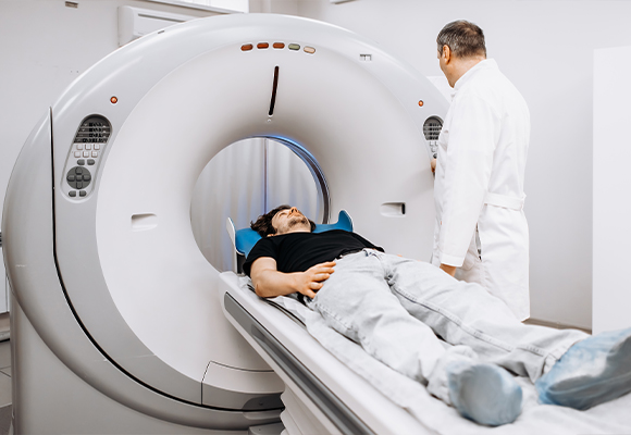
World-class CT Technology

At Ruby Hall Clinic, our commitment to precision and safety is reflected in the advanced CT systems we utilise. These world-class machines are designed to provide detailed diagnostic images and minimal radiation exposure while ensuring ensure the highest standards of accuracy and safety. Our skilled team of radiologists and technicians is dedicated to delivering reliable results while ensuring your comfort every step of the way.

  • Philips Ingenuity Core128 with iDose Technology:
    This 128-slice CT scanner delivers high-resolution images, tailored to individual patient needs, at significantly reduced radiation levels. Its rapid reconstruction capabilities ensure swift and efficient imaging, making it an ideal choice for routine scans and complex angiographies. All procedures are conducted using the safest FDA-approved contrast agents.
  • Siemens Somatom Force VB 30:
    Equipped with dual-source technology, this scanner offers unparalleled accuracy and speed, particularly in cardiac and vascular imaging. Its dual-energy capabilities allow for precise imaging even in challenging clinical scenarios, all while maintaining low radiation doses for patient safety.
 

What is a CT Scan?

A CT (computed tomography) scan is a sophisticated imaging procedure that combines X-rays with advanced computer technology to create cross-sectional views of the body. These highly detailed images enable healthcare professionals to diagnose a wide range of conditions with exceptional clarity.

When is a CT Scan Needed?

CT scans are a crucial diagnostic tool in many areas of medicine, including:

  • Detecting fractures, injuries, or abnormalities in bones and joints.
  • Diagnosing and monitoring conditions such as tumours, infections, or internal bleeding.
  • Evaluating the effectiveness of ongoing treatments, such as chemotherapy or radiation therapy.
  • Planning surgical procedures or minimally invasive interventions.
  • Assessing damage caused by accidents or trauma.


What to Expect During Your CT Scan

Before the Procedure:
Proper preparation is essential to ensure accurate imaging:

  • Please bring all prior medical records, such as X-rays, CT/MRI scans, blood reports and your doctor’s prescription.
  • If a contrast-enhanced scan is required, fasting for at least 4 hours beforehand is necessary. You may continue routine medications and drink plain water during this period.
  • A recent serum creatinine report is mandatory to confirm kidney function before administering contrast material.
  • If you are pregnant, inform the radiologist or technician beforehand, as radiation can pose risks to both mother and baby.
  • Notify the medical team if you have a history of allergies, asthma, or previous reactions to contrast materials.


During the Procedure:

  • You will lie comfortably on a motorised table that moves through the CT scanner, which is shaped like a large ring.
  • Depending on the area being examined, you may be asked to hold your breath briefly to avoid motion blur.
  • If contrast material is needed, it will be administered either orally, intravenously, or rectally, depending on the type of scan.
  • The procedure is painless and typically lasts 5–30 minutes.


Contrast Material Administration

Contrast materials enhance the visibility of specific organs and structures, making it easier to detect abnormalities. They are administered in one of the following ways:

  • Orally: To highlight the stomach and digestive system.
  • Intravenously: For improved imaging of blood vessels, soft tissues and internal organs.
  • Rectally: For detailed views of the lower digestive tract.

While contrast materials are generally well-tolerated, you might experience a brief sensation of warmth or a metallic taste when injected. Our team will closely monitor you throughout the procedure to ensure your comfort and safety.

After the Procedure:

  • You can resume normal activities unless otherwise advised.
  • If contrast material was used, it is recommended to drink plenty of water to aid its removal from your system.
  • Inform the medical team if you notice any unusual symptoms, such as a rash or difficulty breathing.

Understanding Your Results

The images obtained during your scan will be analysed by our expert radiologists, who will prepare a comprehensive report. The findings will be shared with your referring doctor, usually within 24–48 hours. In urgent cases, results are expedited to facilitate timely medical care.

  • Your doctor will use the results to guide your treatment or determine the need for additional tests.
  • If necessary, a digital copy or CD of your scan can be provided for future reference.


Guidelines for Patients

  • Please arrive 15 minutes prior to your scheduled appointment to complete the registration process.
  • Ensure you are accompanied by an attendant for the procedure.
  • Avoid bringing valuables to the hospital.
  • If there are delays, cancellations, or changes to your appointment, please contact us at                 020-6645 5348 / 020-6645 5508.

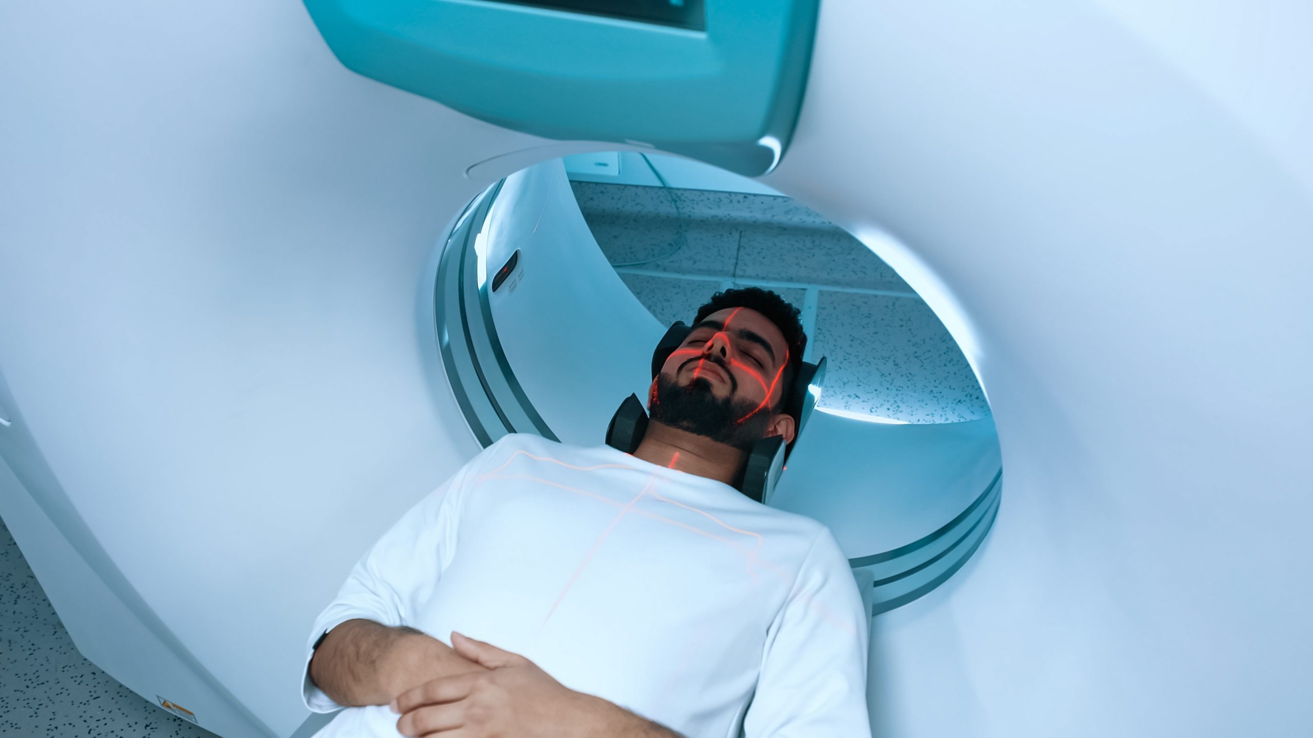
Cutting-Edge MRI Technology

At Ruby Hall Clinic, our dedication to precision and patient safety is reflected in our cutting-edge MRI systems. These state-of-the-art machines provide high-resolution diagnostic images without radiation exposure, ensuring the highest standards of accuracy, safety and patient comfort. Our expert team of radiologists and technicians is committed to delivering reliable results with a seamless and reassuring experience.

  • 3T Philips Ingenia Evolution MRI with SmartSpeed Technology:
    The 3T Philips Ingenia Evolution MRI with SmartSpeed Technology is a state-of-the-art imaging system designed for precision, speed and patient comfort. It delivers unmatched image clarity, ensuring accurate diagnosis of neurological, musculoskeletal and abdominal conditions. SmartSpeed technology enables faster scan times while minimising motion artefacts, improving overall efficiency. The wide-bore design enhances comfort, particularly for patients with claustrophobia, while silent scanning technology provides a noise-reduced, stress-free experience. Additionally, AI-assisted imaging optimises accuracy, making this MRI system a benchmark in advanced diagnostic care.
 

What is an MRI Scan?

Magnetic Resonance Imaging (MRI) is an advanced imaging technique that uses powerful magnetic fields and radio waves to generate high-resolution images of the body’s internal structures. Unlike CT scans, MRI does not use radiation, making it a safe and effective choice for imaging soft tissues, organs and the nervous system.

When is an MRI Scan Needed?

MRI scans are widely used in diagnosing and monitoring a variety of conditions, including:

  • Neurological disorders, such as brain tumours, stroke and multiple sclerosis
  • Spinal conditions, including herniated discs and spinal cord injuries
  • Joint and musculoskeletal injuries, such as ligament tears or cartilage damage
  • Abdominal and pelvic conditions affecting the liver, kidneys, or reproductive organs
  • Cardiac imaging to assess heart structure and function
  • Oncology cases for detecting, staging and monitoring tumours


Risks

MRI is generally a safe and non-invasive procedure, but certain risks should be considered:

  • Metallic Implants – Patients with pacemakers, cochlear implants, aneurysm clips, or certain metal implants may not be suitable for MRI scans due to the strong magnetic field.
  • Contrast Reactions – While contrast agents used in MRI are usually safe, some individuals may experience mild side effects such as nausea, headaches, or allergic reactions. In rare cases, kidney problems can occur, particularly in patients with pre-existing renal conditions.
  • Claustrophobia and Anxiety – Some patients may feel uncomfortable in the enclosed MRI scanner. Our wide-bore design helps alleviate claustrophobia and sedation can be provided if needed.
  • Pregnancy Considerations – While MRI is considered safe during pregnancy, it is typically avoided in the first trimester unless medically necessary. Contrast agents are generally not recommended for pregnant women.
  • Thermal Effects – Prolonged exposure to high-intensity MRI scans may cause slight heating of body tissues, though this is rare and closely monitored.
 

What to Expect During Your MRI Scan

Before the Procedure:
Proper preparation is essential to ensure accurate imaging:

  • Wear comfortable, loose-fitting clothing without metal fasteners or jewellery.
  • Inform the medical team if you have metal implants, pacemakers, or claustrophobia.
  • In some cases, contrast material may be used to enhance image clarity. If required, you may need a recent serum creatinine test to assess kidney function.
 

During the Procedure:

  • You will lie on a cushioned table that slides into the MRI scanner.
  • The scan is painless, but you may hear rhythmic tapping or buzzing sounds from the machine. Our silent scanning technology significantly reduces noise for a more comfortable experience.
  • You may be asked to remain still or briefly hold your breath for clearer images.
  • The procedure typically lasts between 20 to 60 minutes, depending on the area being examined.
 

After the Procedure:

  • You can resume normal activities immediately unless sedation was used.
  • If contrast material was administered, drink plenty of water to help flush it out of your system.
  • Our expert radiologists will analyse your scan and share the results with your referring doctor within 24–48 hours. Urgent cases are prioritised for faster reporting.
 

Guidelines for Patients

  • Please arrive 15 minutes prior to your scheduled appointment to complete the registration process.
  • Ensure you are accompanied by an attendant for the procedure.
  • Avoid bringing valuables to the hospital.
  • If there are delays, cancellations, or changes to your appointment, please contact us at                 020-6645 5348 / 020-6645 5508.

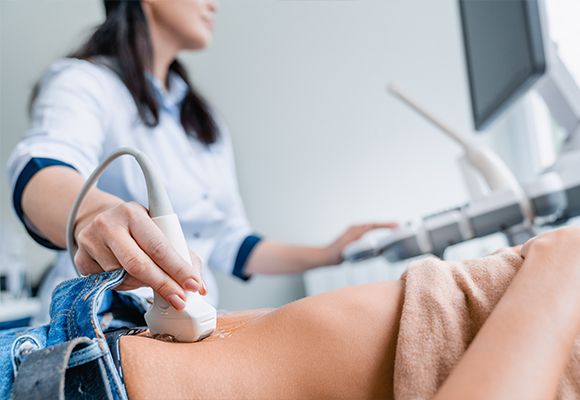
Advanced Ultrasound Technology at Ruby Hall Clinic

At Ruby Hall Clinic, we offer cutting-edge ultrasound imaging designed to provide real-time, high-resolution visuals of internal organs, tissues and blood flow. Our department is equipped with the latest in ultrasound technology, including 3D Colour Doppler, high-frequency probes and portable ultrasonography—ensuring detailed and accurate diagnostics across a wide range of medical specialities.

Whether for routine wellness checks, obstetric evaluations, or guided interventional procedures, our skilled team of radiologists and sonographers delivers precise results with compassion and professionalism.

 What is an Ultrasound Scan?

Ultrasound, also known as sonography, is a non-invasive imaging technique that uses high-frequency sound waves to create real-time images of the body’s internal structures. Unlike X-rays and CT scans, ultrasound does not use radiation, making it a safe and effective diagnostic tool for patients of all ages, including pregnant women and children.

When is an Ultrasound Scan Needed?

Ultrasound is used in a variety of medical applications, including:

  • Abdominal Imaging: Evaluating organs such as the liver, gallbladder, pancreas, kidneys and spleen
  • Obstetric and Foetal Medicine: Monitoring pregnancy, foetal growth and potential complications
  • Breast Imaging and Elastography: Real-time visualisation of breast tissue for cysts, lumps and abnormalities
  • Liver Elastography: Assessing liver stiffness and fibrosis non-invasively
  • Vascular and Colour Doppler Studies: Measuring blood flow in arteries and veins, identifying clots, blockages and vascular disorders
  • Musculoskeletal and Soft Tissue Scans: Diagnosing ligament injuries, joint inflammation and soft tissue concerns
  • Transvaginal and Transrectal Ultrasound: Providing enhanced views for pelvic and prostate evaluations
  • Whole-Body Sonography: Offering an in-depth general assessment when needed
  • USG-Guided Interventions: Including biopsies, fluid drainage and pain management injections, all performed under image guidance for safety and precision
  • Portable and Intraoperative Ultrasound: For use in critical care settings such as the ICU, trauma cases and operating theatres
 

What to Expect During Your Ultrasound

Before the Procedure:
Proper preparation is essential to ensure accurate imaging:

  • Please bring all previous medical records, including X-rays, CT/MRI scans and your doctor’s prescription.
  • For abdominal and pelvic scans: A full bladder is required, so you may be asked to drink water before your appointment.
  • For fasting ultrasounds (e.g., liver, gallbladder): You may be required to avoid food and drink for a few hours before the scan
  • Please inform the medical team if you are pregnant or have any pre-existing medical conditions.
 

During the Procedure:

  • You will lie on an examination table while a trained sonographer applies a water-based gel to the area being examined.
  • A small handheld device called a transducer is gently moved over your skin, emitting sound waves that create live images on a monitor.
  • Depending on the scan type, you may be asked to change positions or hold your breath briefly to enhance image clarity.
  • The procedure is completely painless and typically lasts 15–45 minutes, depending on the complexity of the examination.
 

After the Procedure:

  • You can resume normal activities immediately after your ultrasound.
  • If a biopsy or intervention was performed, follow any post-procedure care instructions provided by your doctor.
  • Drinking plenty of fluids can help if you underwent a Doppler or contrast-enhanced ultrasound.


Understanding Your Results

Our expert radiologists will analyse the ultrasound images and prepare a detailed report for your referring doctor. In most cases, results are available within 24 – 48 hours and urgent cases are prioritised for immediate assessment.

Guidelines for Patients

  • Please arrive 15 minutes prior to your scheduled appointment to complete the registration process.
  • Avoid bringing valuables to the hospital.
  • If there are delays, cancellations, or changes to your appointment, please contact us at 020-6645 5348 / 020-6645 5508.

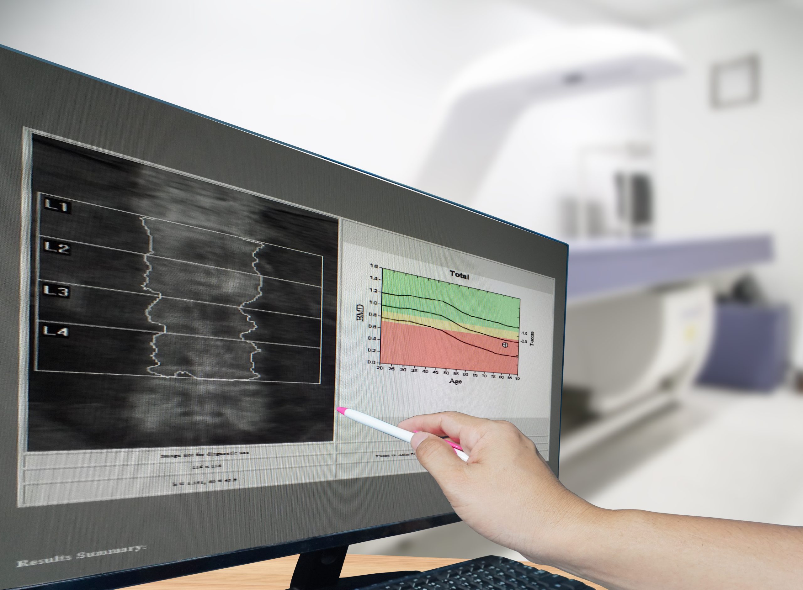
Assessing Bone Health with Precision

At Ruby Hall Clinic, we offer advanced bone density scanning using DEXA (Dual-Energy X-ray Absorptiometry) technology—an essential tool in the early detection and management of osteoporosis and related bone conditions. This safe, low-radiation test provides accurate measurements of bone mineral density (BMD), enabling clinicians to assess fracture risk and monitor treatment effectiveness with precision.

What is a Bone Density Scan?

A bone density scan is a quick, non-invasive test that measures the strength and density of bones, typically in the spine, hip or forearm. It uses two low-dose X-ray beams to calculate how much calcium and other minerals are present in a given segment of bone. The test is completely painless and usually takes less than 20 minutes.

When is a Bone Density Scan Recommended?

Your doctor may recommend a DEXA scan if you:

  • Are a woman over the age of 65 or a man over 70
  • Have a family history of osteoporosis or fractures
  • Have experienced height loss, a stooped posture, or fractures from minor falls
  • Are on long-term corticosteroid therapy or medications affecting bone metabolism
  • Have certain medical conditions such as thyroid disease, rheumatoid arthritis, or chronic kidney disease
  • Are undergoing cancer treatment or early menopause


What to Expect During Your Test:

  • The scan is quick, painless and non-invasive, typically lasting about 15–20 minutes.
  • You will lie on a cushioned table while a scanning arm passes over your body, focusing primarily on the spine and hips.
  • You can wear comfortable clothing, but metal fastenings or jewellery around the scanned areas may need to be removed.
 

Before the Test:

  • No special preparation is usually required.
  • Avoid calcium supplements 24 hours before the scan, unless advised otherwise.
  • Inform the radiology team if you are pregnant or may be pregnant, as a precautionary measure.


Understanding Your Results

The results are reported as T-scores and Z-scores, comparing your bone density to that of a healthy young adult and to someone your age, respectively.

  • A T-score above -1 is considered normal.
  • A score between -1 and -2.5 indicates osteopenia (low bone mass).
  • A T-score below -2.5 is diagnostic of osteoporosis.
 

Guidelines for Patients

  • Please arrive 15 minutes prior to your scheduled appointment to complete the registration process.
  • Avoid bringing valuables to the hospital.
  • If there are delays, cancellations, or changes to your appointment, please contact us at 020-6645 5348 / 020-6645 5508.

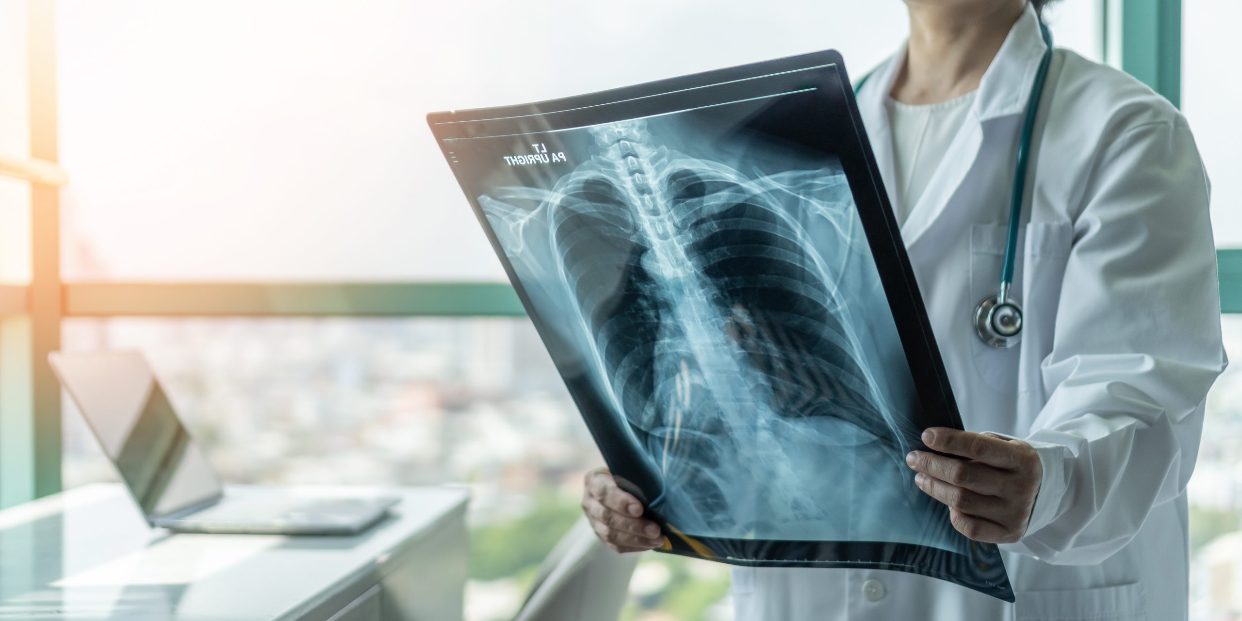
At Ruby Hall Clinic, our X-ray facilities combine precision, speed and safety to deliver high-quality diagnostic imaging. As one of the most commonly used medical imaging techniques, X-rays allow our skilled radiologists to assess bones, joints and soft tissues with remarkable clarity. We use advanced digital X-ray technology, ensuring minimal radiation exposure while providing accurate and timely diagnoses.

Shimadzu Flexavision F3 – Fully Digital R/F Mobile X-ray System

The DRX is a filly digital system with portable wireless dynamic flat-panel detectors (FPD) for routine and emergency radiology with lowest radiation. The FPD provides a large field of view capable of fluoroscopic applications.The system supports a wide range of examinations, from barium enema to gastrointestinal, non-vascular interventional radiology procedures, DIP and other urinary tract contrast media acquisitions.

What is an X-Ray?

An X-ray is a non-invasive imaging technique that uses a small dose of ionising radiation to capture images of the body’s internal structures. This quick and painless procedure is essential for detecting fractures, infections, lung conditions and various other medical concerns.

Why is an X-Ray Done?

X-rays are used to diagnose a range of conditions, including:

  • Bone Fractures and Joint Injuries – Helps identify broken bones, dislocations and arthritis-related changes.
  • Lung and Chest Conditions – Detects pneumonia, tuberculosis, lung tumours, or fluid accumulation.
  • Dental and Jaw Issues – Assesses cavities, impacted teeth and jawbone abnormalities.
  • Abdominal and Digestive Concerns – Identifies swallowed objects, kidney stones, or intestinal blockages.
  • Spinal and Skeletal Disorders – Evaluates scoliosis, osteoporosis, or other degenerative conditions.


Before the Procedure:
Proper preparation is essential to ensure accurate imaging:

  • You may be asked to remove any jewellery, metal accessories, or clothing with zippers or buttons that could interfere with the images.
  • If you are pregnant or suspect pregnancy, inform our radiology team beforehand to discuss necessary precautions.
 

During the Procedure:

  • Our radiology technician will position you carefully to ensure the best possible image quality.
  • You may be asked to hold your breath and remain still for a few seconds while the X-ray is taken.
  • The machine emits a brief, painless pulse of radiation to capture the image.
  • Depending on the type of X-ray, the process may take anywhere from a few minutes to 30 minutes.


After the Procedure:

  • In most cases, you can resume normal activities immediately after the scan.
  • If a contrast agent was used, drinking plenty of water will help flush it out of your system.
  • Our expert radiologists will analyse the X-ray results and share them with your doctor promptly for further evaluation.
 

Safety and Radiation Exposure

At Ruby Hall Clinic, patient safety is our top priority. While X-rays involve exposure to a small amount of radiation, our advanced digital imaging technology ensures that doses are kept to an absolute minimum.

  • Low-Dose Radiation Technology – Our systems are designed to use the lowest effective radiation levels while maintaining image quality.
  • Shielding Measures – Protective lead aprons and collars are provided when necessary to minimise exposure.
  • Special Precautions for Pregnant Women and Children – Alternative imaging methods may be recommended when possible to avoid unnecessary radiation.

Thanks to these advancements, X-rays remain one of the safest and most efficient diagnostic tools available.

Guidelines for Patients

  • Please arrive 15 minutes prior to your scheduled appointment to complete the registration process.
  • Avoid bringing valuables to the hospital.
  • If there are delays, cancellations, or changes to your appointment, please contact us at 020-6645 5348 / 020-6645 5508.

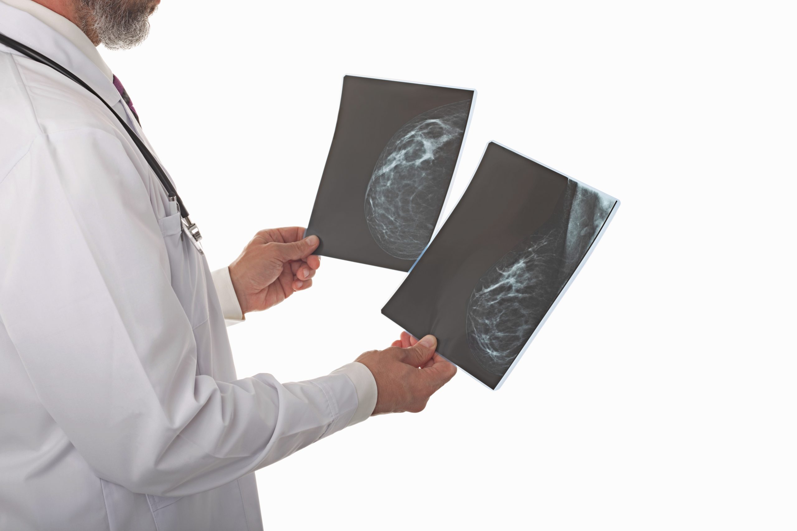
Advanced Breast Imaging for Early Detection

At Ruby Hall Clinic, we understand the importance of early detection when it comes to breast health. That’s why our Mammography Services are designed to combine advanced technology with expert care in a compassionate and reassuring environment. Whether for routine screening or diagnostic evaluation, our all-women team of radiologists, sonologists, breast surgeons, radiation oncologists and genetic counselling experts ensures a seamless, personalised experience tailored to each patient.

GE Pristina Digital 3D Tomosynthesis

Ruby Hall Clinic is proud to be the first in Asia to offer the GE Senographe Pristina Digital 3D Mammography system—a breakthrough in patient-centric breast imaging. What sets this system apart?

Self-Compression Tool: Unique to the Pristina platform, this feature empowers women to control the compression during their mammogram, enhancing comfort and reducing anxiety.

High-Resolution 3D Imaging: Enables clearer, more accurate views of breast tissue, especially beneficial for women with dense breasts.

Ergonomic Design: Built with women in mind, reducing physical strain and allowing easier, more precise positioning for both patient and technologist.

Comprehensive Support: The dedicated breast imaging suite is operated by a highly skilled and empathetic female-led team, ensuring privacy and comfort at every step.

What is a Mammogram?

A mammogram is a specialised X-ray examination of the breast used to detect and evaluate changes in breast tissue. It plays a crucial role in the early diagnosis of breast cancer, often identifying abnormalities before symptoms appear. Mammography can detect tumours that are too small to be felt and help distinguish between benign and suspicious findings.

There are two main types:

  • Screening mammograms: For women without symptoms, usually performed as part of routine health checks.
  • Diagnostic mammograms: For investigating specific concerns such as a lump, pain, nipple discharge, or skin changes.


When is Mammography Recommended?

Mammography is typically advised:

  • Annually or biennially for women aged 40 and above, depending on individual risk factors.
  • Earlier or more frequently for those with a family history of breast cancer or other high-risk indicators.
  • As a follow-up to clinical breast exams or when symptoms such as lumps, pain, or skin changes are present.


What to Expect During the Procedure?

  • You will be asked to undress from the waist up and wear a provided gown.
  • Each breast is positioned on a flat surface and gently compressed for a few seconds while images are taken from different angles.
  • The procedure may cause mild discomfort but is typically brief and well tolerated
  • The entire exam usually takes about 20 minutes.


After the Procedure:

  • Your images are reviewed by our specialist radiologists and a detailed report is sent to your referring doctor as well as handed over to you.
  • In some cases, additional imaging such as ultrasound or MRI may be recommended for further evaluation.
 

Guidelines for Patients

  • Please arrive 15 minutes prior to your scheduled appointment to complete the registration process.
  • Avoid bringing valuables to the hospital.
  • If there are delays, cancellations, or changes to your appointment, please contact us at 020-6645 5348 / 020-6645 5508.

Minimally Invasive, Targeted Treatment

Ruby Hall Clinic’s Interventional Radiology services are led by a team of highly skilled specialists, supported by state-of-the-art imaging systems such as the Philips Ingenuity Core128 and Siemens Somatom Force VB 30, ensuring accuracy, safety and comfort for every procedure.

What is Interventional Radiology?

Interventional Radiology (IR) combines the precision of advanced imaging with minimally invasive techniques to diagnose and treat various medical conditions. Using real-time guidance from CT or ultrasound, our radiologists can access even the most delicate areas of the body, offering an alternative to conventional surgery with shorter recovery times and fewer complications.

When Is Interventional Radiology Needed?

Interventional Radiology is typically recommended when traditional diagnostic methods or surgical interventions are either too invasive, carry higher risk, or are not suitable for the patient due to underlying health conditions. It plays a crucial role in:

  • Diagnosing suspicious growths or lesions through image-guided biopsies or FNAC
  • Managing chronic or localised pain when medication and physical therapy prove ineffective
  • Draining abnormal fluid collections in the chest or abdomen (pleural effusions or ascites)
  • Providing treatment for small tumours using minimally invasive techniques like radiofrequency ablation
  • Evaluating and addressing spinal or neurological concerns where MRI is contraindicated or inconclusive
  • Tracing abnormal tracts or sinuses for surgical planning
  • Guiding therapy with real-time imaging to ensure accurate targeting and minimal collateral impact
 

Whether for diagnosis or treatment, IR is often the first step towards a more accurate, safer and faster road to recovery. At Ruby Hall Clinic, our team ensures that every procedure is thoughtfully chosen and meticulously performed, with the patient’s comfort and outcome as the highest priorities.

Types of Procedures

CT Guided Procedures

  • CT-Guided Biopsy
    This procedure involves extracting a small tissue sample from an organ or suspected lesion using a fine needle, guided by CT imaging. It enables accurate diagnosis of conditions such as tumours, infections, or inflammatory diseases—without the need for open surgery.
  • CT-Guided FNAC (Fine Needle Aspiration Cytology)
    FNAC is used to sample fluid or cells from abnormal masses for cytological examination. With the help of CT guidance, the needle is placed precisely, even in deep-seated or difficult-to-access areas, ensuring safe and conclusive results.
  • CT-Guided Pain Block
    This targeted procedure delivers medication directly to nerves or joints that are the source of chronic pain. Using CT imaging ensures accurate placement of the injection, offering effective relief in conditions like spine or joint disorders, especially when other treatments have failed.
  • CT-Guided Radiofrequency Ablation (RFA)
    RFA is a minimally invasive technique used to destroy abnormal tissues, such as tumours, using heat generated by radiofrequency energy. Under CT guidance, a needle electrode is positioned precisely within the target area. The technique is often used in treating liver, kidney, lung and bone lesions.


Spinal and Neurological Imaging

  • Cisternography
    This specialised scan evaluates the flow of cerebrospinal fluid (CSF) in the brain and spinal canal. It is helpful in diagnosing CSF leaks, hydrocephalus, or intracranial pressure issues. A contrast agent is introduced into the CSF space and images are taken to assess any abnormalities.
  • Myelography
    Used to detect problems in the spinal cord, nerve roots, or other spinal structures, myelography involves injecting contrast material into the spinal canal. This test is particularly useful in diagnosing spinal stenosis, herniated discs, or tumours when MRI is inconclusive or contraindicated.
 

Contrast Studies and Track Visualisation

  • Sinogram
    A sinogram visualises abnormal sinus tracts or fistulas. It involves injecting a contrast dye into the external opening of a sinus tract and capturing detailed images to trace its path. This helps determine the extent and connections of the tract, which is essential before surgical planning.
 

Ultrasound-Guided Procedures

  • USG-Guided Biopsy
    Using real-time ultrasound imaging, a needle is directed to the area of interest—such as the liver, thyroid, or lymph nodes—to extract tissue samples. This method is quick, accurate and ideal for soft-tissue masses that are visible on ultrasound.
  • USG-Guided FNAC
    Similar to biopsy but using a thinner needle, FNAC under ultrasound guidance is employed to obtain cellular material from lumps or fluid collections for diagnostic purposes. It’s commonly performed on thyroid nodules, breast lumps, or enlarged lymph nodes.
  • USG-Guided Pleural / Ascitic Tapping
    This procedure involves the safe drainage of excess fluid from the pleural cavity (around the lungs) or the peritoneal cavity (in the abdomen) under ultrasound guidance. It provides symptomatic relief and allows for fluid analysis to determine the underlying cause of fluid accumulation.
 

What to Expect for the Procedure?

Before the Procedure:

  • Please carry prior imaging reports, doctor’s prescriptions and recent blood tests.
  • Some procedures may require fasting or pre-procedure blood work.
  • Inform the radiologist if you have bleeding disorders, allergies (especially to contrast material), or if you’re pregnant.
  • You will be asked to sign a consent form before the procedure.
 

During the Procedure:

  • Most procedures are performed under local anaesthesia with real-time imaging guidance.
  • You may feel slight pressure or discomfort, but pain is minimal.
  • The radiologist and nurse will monitor you throughout for safety and comfort.
 

After the Procedure:

  • You may need to rest for a short period post-procedure.
  • In most cases, patients return home the same day.
  • Post-procedural care instructions will be provided, including signs to watch for.
  • Results are typically shared with the referring doctor within 24–48 hours.
 

Guidelines for Patients

  • Please arrive 15 minutes prior to your scheduled appointment to complete the registration process.
  • Avoid bringing valuables to the hospital.
  • If there are delays, cancellations, or changes to your appointment, please contact us at 020-6645 5348 / 020-6645 5508.

Precision. Preservation. Peace of Mind.

At Ruby Hall Clinic, our comprehensive suite of image-guided breast interventions is designed to detect abnormalities early, guide treatment effectively and minimise trauma to healthy tissue. From biopsies and wire localisation to vacuum-assisted scarless surgery, every procedure is conducted with the utmost attention to detail, safety and patient comfort.

What are Breast Interventions?

Breast interventions at Ruby Hall Clinic offer a sophisticated blend of diagnostic accuracy and therapeutic precision, tailored specifically for breast-related conditions. Using state-of-the-art imaging guidance — including ultrasound, mammography and stereotactic systems — we perform targeted procedures that eliminate the need for extensive surgery in many cases.

When Are Breast Interventions Needed?

Breast interventions are recommended when:

  • Suspicious lesions or abnormalities are detected on a mammogram, ultrasound, or MRI
  • Microcalcifications are observed that require further investigation through stereotactic biopsy
  • Benign conditions, such as fibroadenomas or breast cysts, cause discomfort or anxiety and need removal or aspiration
  • Infections or collections like abscesses, seromas, or post-operative fluid require drainage
  • Granulomatous mastitis, a rare inflammatory breast condition, needs precise steroid therapy
  • Pre-surgical planning demands wire localisation or marker clip placement to accurately identify areas for excision
  • Lymph nodes in the axilla (armpit) appear suspicious and need tissue sampling for staging or diagnosis
 

These interventions are minimally invasive, often done under local anaesthesia and allow for faster recovery, better cosmetic outcomes and accurate pathology reports to guide the next steps in care.

Types of Breast Interventions Offered

  • Image-Guided Core Needle Biopsy
    Performed under ultrasound or mammographic guidance, this procedure removes small tissue samples from a suspicious breast lesion for detailed histological evaluation.
  • Stereotactic Biopsy for Microcalcifications
    Recommended when tiny calcium deposits are detected on mammograms. Using a special mammography unit, the area is precisely targeted for biopsy.
  • Vacuum-Assisted Fibroadenoma Resection (VAB)
    A scarless, minimally invasive alternative to surgery for benign breast lumps such as fibroadenomas. It offers excellent cosmetic results and is performed under imaging guidance.
  • Wire Localisation
    Used prior to breast surgery, a thin wire is placed to mark the exact location of a lesion that is not felt but seen on imaging — helping the surgeon excise it accurately.
  • Clip Placement
    A tiny marker clip is placed at the site of a biopsy-proven lesion, particularly useful for follow-up or guiding future surgery or treatment.
  • Aspirations for Abscess / Cyst / Seroma
    Fluid or pus collections are aspirated under ultrasound guidance to relieve discomfort, confirm diagnosis, or prevent infection.
  • Axillary Node FNAC / Biopsy
    Performed when lymph nodes in the axilla show abnormal size or features. Helps in cancer staging or ruling out metastasis.
  • Kenacort Injections for Idiopathic Granulomatous Mastitis (IGM)
    A targeted steroid injection used to treat this rare, chronic inflammatory breast condition, avoiding systemic side effects.
  • Scarless Surgery for Benign Breast Lesion
    A minimally invasive, image-guided procedure that removes lumps like fibroadenomas through a tiny incision, offering excellent cosmetic results without stitches or visible scarring.
 

What to Expect During the Procedure?

  • Most breast interventions are day-care procedures, performed under local anaesthesia with little to no downtime.
  • A specialist radiologist will explain the steps, address your concerns and ensure optimal imaging is used to guide the procedure.
  • You may feel mild pressure or discomfort during certain procedures, but pain is minimal.
 

After the Procedure:

  • Tissue samples collected are sent to pathology for detailed examination. Reports typically take 3–5 working days.
  • These results help determine if further treatment, surgery, or monitoring is required.
  • If a clip has been placed, patients are advised to inform their doctor and future imaging teams.
  • Follow-up imaging or appointments may be scheduled depending on findings.
 

Guidelines for Patients

  • Please arrive 15 minutes prior to your scheduled appointment to complete the registration process.
  • Avoid bringing valuables to the hospital.
  • If there are delays, cancellations, or changes to your appointment, please contact us at 020-6645 5348 / 020-6645 5508.

Meet Our Doctors

Dr. Pranav Mahadeokar

Head, Department of Radiology

Dr. Tanvi Vaidya

Division of GI Radiology

Dr. Yadav Munde

Interventional Radiology

Dr. M. K. Mutatkar

Interventional Radiology

Dr. Rochan Pant

Interventional Radiology

Dr. Gurtej Singh

Interventional Radiology